Back in April we launched this year’s breast cancer research image competition. The competition was open to anyone affiliated with a research institution or hospital and entrants were asked to submit images that describe some aspect of breast cancer research, with categories ranging from images taken down the microscope, computer generated creations and life in the lab or clinic.
Thank you to everyone who entered. Picking the overall winner was no easy task for the Breast Cancer Now Scientific Advisory Board and there were some really stunning entries.
We’d like to say a huge congratulations to the Overall Winner, Ansel Lim and the Supporters’ Choice Winner Sarah Boyle.
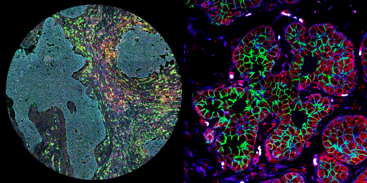
Overall Winner – Rainbow Kaleidoscope by Ansel Lim
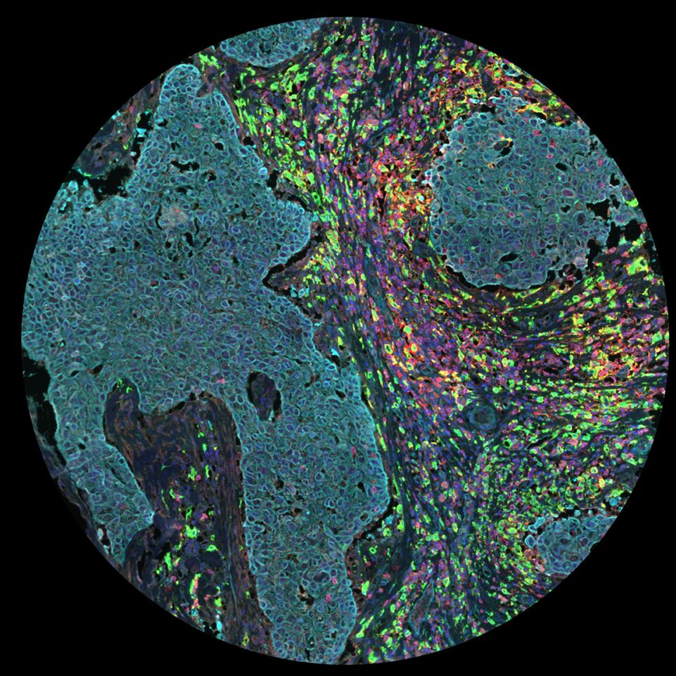
About the image: This stunning image showcases an unwitting accomplice in the spread of one of the most aggressive types of breast cancer. Taking center stage in this patient specimen is what we call an “M2 macrophage”. That’s the red and green cell you see smack in the middle of this colorfully stained slide, floats aimlessly around islands of cancer in an ocean of a thousand defending immune cells. We identikit it with the colors of the rainbow. This “special snowflake” sits at the crossroads of an epic battle, one between alien cancer cells and an entire battalion of immune cells fighting off these intruders. This soldier is a Trojan horse. It should have been fighting the enemy, but instead it has been hijacked. A conspiracy! Manipulated by a complex network of signals, it now works for the enemy. It promotes growth of the cancer as it invades other parts of our body. But its attempt at subverting our body is no longer a secret. We can see the cell in plain sight. It can hide no longer from our microscopes. Empowered by the latest technology, doctors and biologists at our hospital work hand in hand to unravel the mysteries of the M2 macrophage.
Supporters’ Choice Winner – Neoplastic Petals by Sarah Boyle
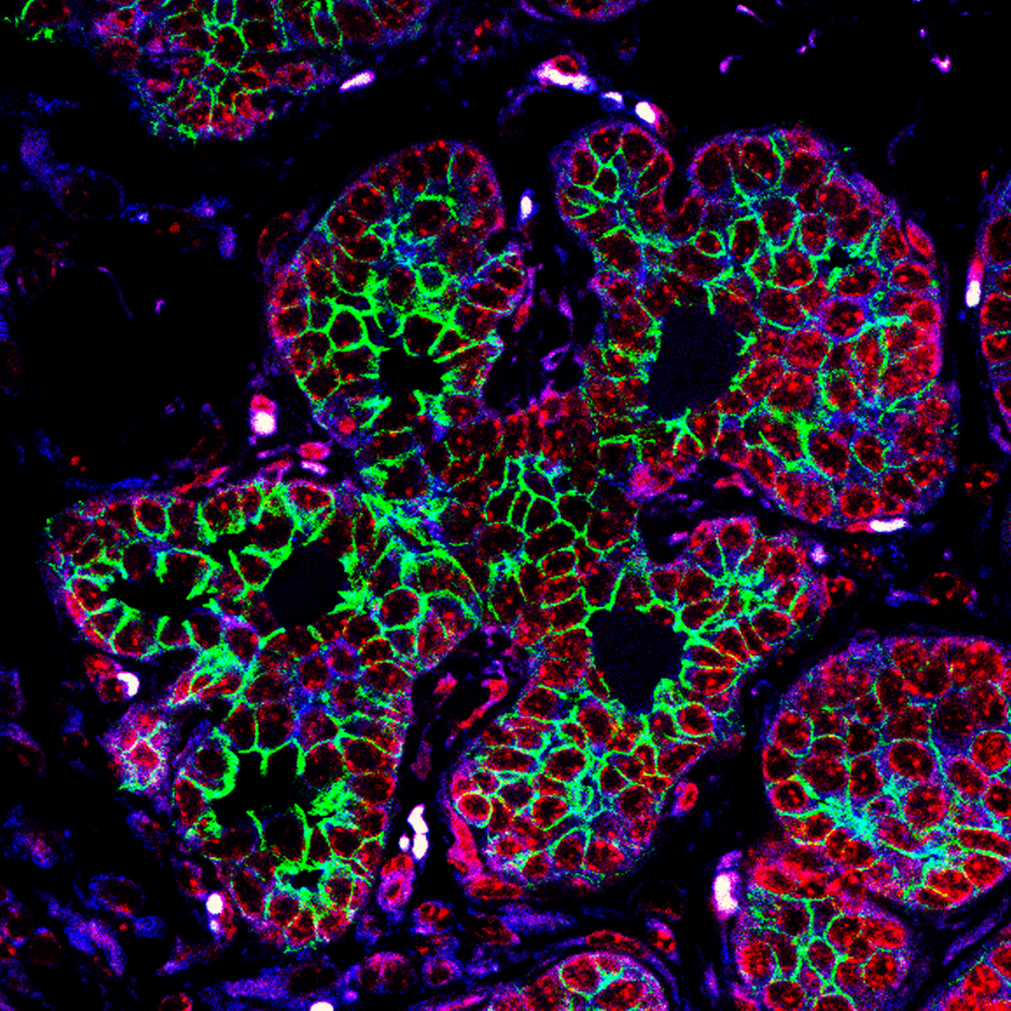
About the image: This is an image of the early stages of cancer within a mammary gland of a genetically-engineered mouse. The green colour labels a protein that is involved in controlling cell movement. As the tumour grows, cells begin to become more mobile, which can lead to cancer spread. Junctions between cells are labelled in blue and cell nuclei in red.
Other images we liked
Sophie Roberts, University of Leeds
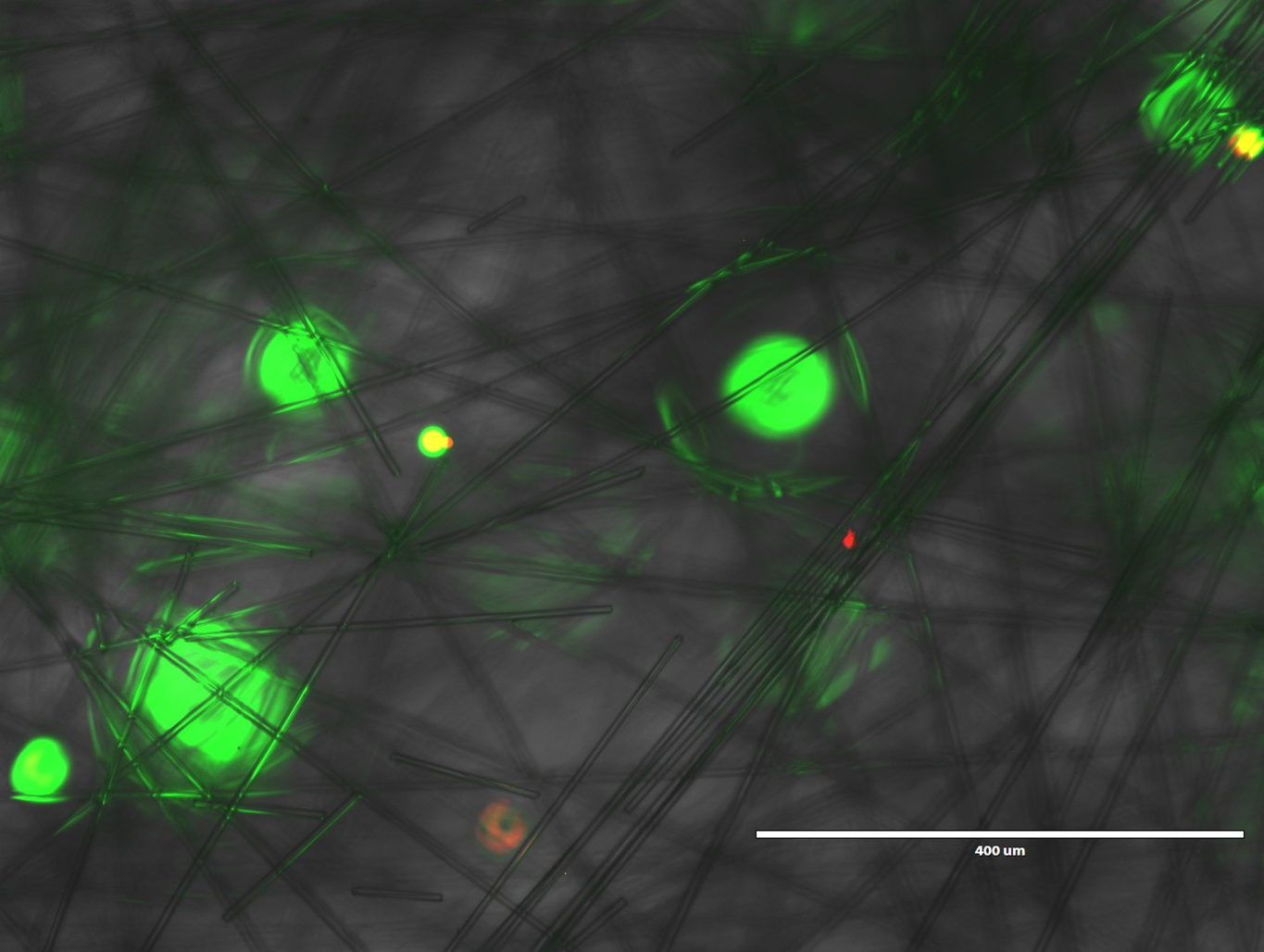
The image shows breast cancer cells that have been cultured in 3D. By culturing them in 3D they more realistically mimic the characteristics of a human breast tumour. The cells are coloured using a stain which stains cells that are alive green and those that are dead in red. It is hoped that by creating a 3D model of breast cancer the use of animals in preclinical drug testing can be reduced.
Matthew Humphries, Univerity of Leeds
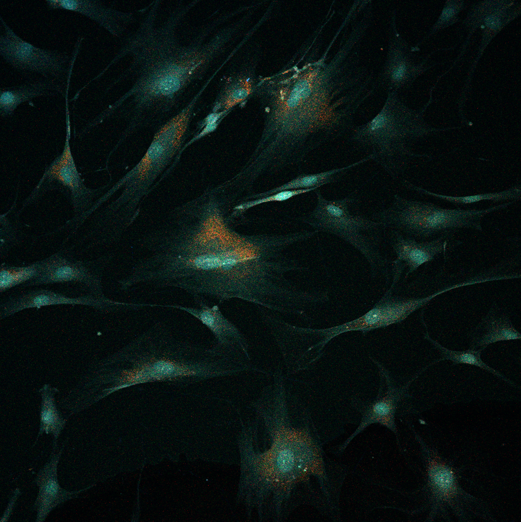
The image is a confocal 2D capture of male breast cancer associated fibroblasts stained with MMP9 (red), B-Actin (green) and DAPI (blue).
Sodium current traces by Theresa Leslie, University of York
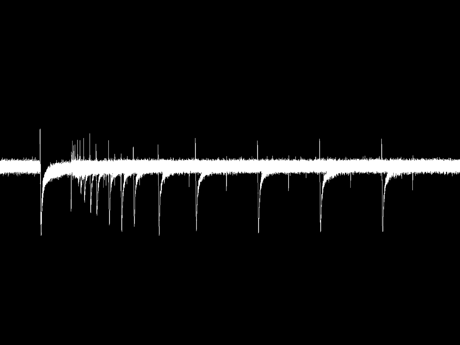
Breast cancer cells are electrically active, and they may use this property to communicate with their environment and invade into different areas of the body. Ions can flow through channels in the cell membrane, causing tiny electrical currents. Drugs can target these ion channels to control the electrical signals in cancer cells and this may reduce the spread of cancer. The electrical current across a single cell membrane can be recorded in real time using microscopic glass electrodes and very sensitive amplifiers. Drugs can be added to the cells during recording to assess their effect on the cancer cells. This is an example of some sodium currents in a breast cancer cell. We are also investigating ion channels in live tissue from patients, provided by Breast Cancer Now.
All images have been released under a Creative Commons Attribution License (CC BY), so everyone is welcome and encouraged to share them freely, while attributing the image author.
Comments