Nanoparticles have many unique properties that are inherent based on their small size. They can be taken up into living cells and have a high surface area relative to their volume which allows, for example, for increased interactions with cell structures and relatively large amounts of a drug to be delivered to a particular site/cell.
Another advantage of utilizing nanoparticles is that they can be designed and synthesized to have multiple functions. We have developed a bioluminescent magnetic nanoparticle system that allows for imaging and tracking of cells and also cellular manipulation due to the nanoparticles’ magnetic properties. This multifunctionality could be further increased if targeting or drug moieties were to be added to the nanoparticle.
The challenges to overcome
There are many challenges associated with the bioimaging of live cells particularly when combined with nanoparticles.
There are many challenges associated with the bioimaging of live cells particularly when combined with nanoparticles. Manipulation of the cells during their incubation with the nanoparticles and during the imaging has to be done carefully to avoid negatively impacting cell health.
Length scale differences between the nanoparticles and living cell structures can make it challenging to image specific interactions between the two. And, of course, living cells—especially motile spermatozoa in a liquid medium—are in motion, so imaging them requires specialized equipment and training.
The approach we have designed for bioimaging combines magnetic nanoparticles with luciferase, an enzyme derived from fireflies that produces bioluminescence. This new nanotech imaging method has the potential to replace currently used cadmium-based quantum dot bioimaging tools that have been shown to cause biotoxicity.
In fact, the magnetic nanoparticles used in this study are already approved for clinical use by the European Medicines Agency and the U.S. Food and Drug Administration.
So, why did we do this?
While these bioluminescent nanoparticles can be used with other cells types, there are several reasons we were interested in testing this new tool on spermatozoa.
First, since the nanoparticles are magnetic, once they are bound to cells not only can the cells be imaged and the motion and position be tracked, but they can also be manipulated using an external magnetic field allowing for cellular locomotive manipulation.
In the case of sperm, this type of manipulation of the cells can be applied to increase fertilization rates. In many assisted reproduction technologies, sperm are directly manipulated by placing them near or inside of eggs.
Using a magnetic method provides the potential for a completely non-mechanical positioning of the sperm minimizing cell damage.
Using a magnetic method provides the potential for a completely non-mechanical positioning of the sperm minimizing cell damage. Similarly, other cutting-edge approaches to manipulating sperm motility have included a corkscrew type motor (original paper).
Second, this new method has significant potential to be expanded to allow customization of the nanoparticle surface. For instance, markers can be identified and targeted allowing the nanoparticles to selectively bind low-quality sperm so that they can be easily sorted from healthy sperm.
Future work
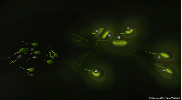
This proof-of-concept work demonstrates nanoparticle binding with animal (boar) spermatozoa with imaging and magnetic function intact after binding. It is demonstrated that these luciferase-modified magnetic nanoparticles can be used effectively for cellular imaging and tracking.
The next steps to advance this work include examining location-specific binding (sperm head vs. the tail), using targeting moieties to control the binding site, controlling nanoparticle density per cell, and targeting specific cell types such as cancer cells.
Certainly, as this technology is further developed, concentration dependent biotoxicity, ultimate particle fate, and impacts due to cellular manipulation from magnetic cellular locomotive manipulation will also be examined.
This nanotechnology platform has significant potential with many avenues of investigation, and we look forward to continued contributions in this challenging and fascinating area of bionanotechnology.
Keisha Walters
Her research covers a broad range of topics in polymer- and nano-based materials engineering and transport modeling.
Latest posts by Keisha Walters (see all)
- Why we created glow in the dark magnetic sperm - 23rd March 2016
That’s incredible. With the ability of manipulation, the tech can help to achieve more. Take the sperm for example. It can assist in the selecting process, first observing the activity of sperms and target one and make it combine with egg.